Upper Back Anatomy : Upper Back Anatomy Artwork Stock Image C014 6967 Science Photo Library / Try the injurymap exercise app now.. Try the injurymap exercise app now. The spine is made up of 33 individual bones called ve. It is very stiff, and the thoracic spine has a limited range of motion. The deltoid, teres major, teres minor, infraspinatus, supraspinatus (not shown) and subscapularis muscles (not shown) all extend from the scapula to the humerus and act on the shoulder joint. The hurt can stem from sore muscles, ligaments, and tendons, or from herniated disks, fractures, and other problems in your upper, middle, and lower back.
In the upper back region the trapezius rhomboid major and levator scapulae muscles anchor the scapula and clavicle to the spines of several vertebrae and the occipital bone of the skull. In the upper back region, the trapezius, rhomboid major, and levator scapulae muscles anchor the scapula and clavicle to the spines of several vertebrae and the occipital bone of the skull. The back functions are many, such as to house and protect the spinal cord, hold the body and head upright, and adjust the movements of the upper and lower limbs. Both the deltoid and the trapezius are firmly attached to the spine of the scapula. The human spine is composed of 4 sections of vertebrae.
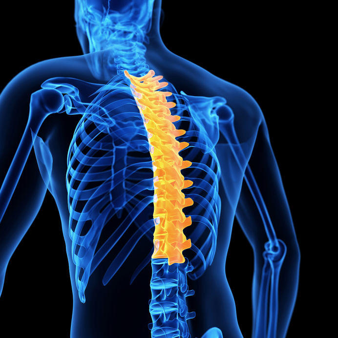
Back Pain Thoracic Joint Restriction from creeksidechiro.com It's hard to underestimate the importance of the spine in your overall anatomy. In order to understand why upper back pain occurs, it's helpful to know the basic anatomy of the spine. The thoracic spine —also referred to as the upper back or middle back—is designed for stability to anchor the rib cage and protect vital internal organs within the chest. The superficial back muscles are situated underneath the skin and superficial fascia. The rib cage also anchors the bones of the head, neck, shoulders, and arms to the trunk of the body. It runs from the neck to the upper back. The spine is made up of 33 individual bones called ve. In the upper back region the trapezius rhomboid major and levator scapulae muscles anchor the scapula and clavicle to the spines of several vertebrae and the occipital bone of the skull.
The spine is made up of 33 individual bones called ve.
Looking for a solution to your back pain problem? These sections are cervical (neck), thoracic (upper and middle back), lumbar (lower back), and sacrum (tailbone). It is very stiff, and the thoracic spine has a limited range of motion. This muscle is located on the upper portion of the back anatomy, underneath the trapezius. It contains many muscles and nerves but only has one bone, the femur, which is the longest and strongest bone in. Upper back pain rear view of spine back pain spine sports spine spine surgery spine white background back ache x human anatomy illustration human anatomy on white background upper body stretch. It's hard to underestimate the importance of the spine in your overall anatomy. In order to understand why upper back pain occurs, it's helpful to know the basic anatomy of the spine. Sometimes you feel the effects right away. The nervous system of the thorax is a vital part of the nervous system as a whole, as it includes the spinal cord, peripheral nerves, and autonomic ganglia that communicate with and control many vital organs. The superficial back muscles are situated underneath the skin and superficial fascia. Upper back pain is most commonly caused by muscle irritation or tension, also called myofascial pain. The thigh bears much of the load of the body's weight when a person is upright.
The spine is made up of 33 individual bones called ve. In the upper back region, the trapezius, rhomboid major, and levator scapulae muscles anchor the scapula and clavicle to the spines of several vertebrae and the occipital bone of the skull. The back functions are many, such as to house and protect the spinal cord, hold the body and head upright, and adjust the movements of the upper and lower limbs. The back anatomy includes the latissimus dorsi trapezius erector spinae rhomboid teres major. Powerful muscles that move the head and arms attach to these bones as well.
Massage For Upper Back Pain Erector Spinae from www.painscience.com Back muscles anatomy here include the trapezius, latissimus dorsi, rhomboid and levator scapulae. Sometimes you feel the effects right away. The spine is made up of 33 individual bones called ve. The upper back originates at the base of your neck, incorporates both shoulders and extends down to mid spine, including your ribs. These include the cervical vertebrae in the neck, the thoracic vertebrae of the ribcage in the upper and middle back, the lumbar vertebrae in the lower back, and the vertebrae that are part of the pelvis. The rib cage also anchors the bones of the head, neck, shoulders, and arms to the trunk of the body. Powerful muscles that move the head and arms attach to these bones as well. These sections are cervical (neck), thoracic (upper and middle back), lumbar (lower back), and sacrum (tailbone).
The muscles of the chest and upper back occupy the thoracic region of the body i.
In the upper back region the trapezius rhomboid major and levator scapulae muscles anchor the scapula and clavicle to the spines of several vertebrae and the occipital bone of the skull. The nervous system of the thorax is a vital part of the nervous system as a whole, as it includes the spinal cord, peripheral nerves, and autonomic ganglia that communicate with and control many vital organs. Powerful muscles that move the head and arms attach to these bones as well. The lumbar and sacrum region make up the bone of the lower back anatomy. The complexity of this region means that dysfunction can occur either due to injury or progressive pain and degeneration. The cause may be poor posture (such as forward head posture) or any type of irritation of the large back and shoulder muscles, including muscle strain or spasms. The basic anatomy of your upper back by lindsey mcfadden as you're doing your regular upper back stretching exercises , you're probably wondering about the components of your upper back and why it happens to be the most stable part of your spine. It contains many muscles and nerves but only has one bone, the femur, which is the longest and strongest bone in. The upper back originates at the base of your neck, incorporates both shoulders and extends down to mid spine, including your ribs. Human body anatomy female female anatomy muscle shoulder blade pain anatomy back muscles bones man female anatomy body muscles in a body female anatomy muscole shoulder concept muscular sysyem. The spine is made up of 33 individual bones called ve. The deltoid, teres major, teres minor, infraspinatus, supraspinatus (not shown) and subscapularis muscles (not shown) all extend from the scapula to the humerus and act on the shoulder joint. The thigh bears much of the load of the body's weight when a person is upright.
The cervical spine supports the weight and movement of your head and protects the nerves exiting your brain. The basic anatomy of your upper back by lindsey mcfadden as you're doing your regular upper back stretching exercises , you're probably wondering about the components of your upper back and why it happens to be the most stable part of your spine. In order to understand why upper back pain occurs, it's helpful to know the basic anatomy of the spine. Both the deltoid and the trapezius are firmly attached to the spine of the scapula. All these muscles are therefore associated with movements of the upper limb.
1 from All these muscles are therefore associated with movements of the upper limb. The iliocostalis muscles are furthest from the spine. It contains many muscles and nerves but only has one bone, the femur, which is the longest and strongest bone in. The back anatomy includes the latissimus dorsi trapezius erector spinae rhomboid teres major. The bones of the chest and upper back combine to form the strong, protective rib cage around the vital thoracic organs such as the heart and lungs. There is a set of muscles in the upper back (called the thoracic area) called the spinalis thoracis. It is key to medicine and other areas of health. In the upper back region, the trapezius, rhomboid major, and levator scapulae muscles anchor the scapula and clavicle to the spines of several vertebrae and the occipital bone of the skull.
The upper back originates at the base of your neck, incorporates both shoulders and extends down to mid spine, including your ribs.
It consists of seven vertebrae. See human back anatomy stock video clips. The rhomboid muscle is activated as you bring and squeeze your scapula or shoulder blades back and together. The rib cage also anchors the bones of the head, neck, shoulders, and arms to the trunk of the body. The lumbar and sacrum region make up the bone of the lower back anatomy. The basic anatomy of your upper back by lindsey mcfadden as you're doing your regular upper back stretching exercises , you're probably wondering about the components of your upper back and why it happens to be the most stable part of your spine. The cervical spine protects the nerves connecting to. The thoracic spine —also referred to as the upper back or middle back—is designed for stability to anchor the rib cage and protect vital internal organs within the chest. Upper back pain is most commonly caused by muscle irritation or tension, also called myofascial pain. Sometimes you feel the effects right away. These include the cervical vertebrae in the neck, the thoracic vertebrae of the ribcage in the upper and middle back, the lumbar vertebrae in the lower back, and the vertebrae that are part of the pelvis. The muscles of the chest and upper back occupy the thoracic region of the body i. Both the deltoid and the trapezius are firmly attached to the spine of the scapula.


Posting Komentar
0 Komentar